
HE stain is routine stain. Histological staining is a vital step in diagnosing various diseases and has been used for more than a century to provide contrast in tissue sections rendering the tissue constituents visible for.

Lesson 12 Metachromatic Staining Pdf Staining Histology
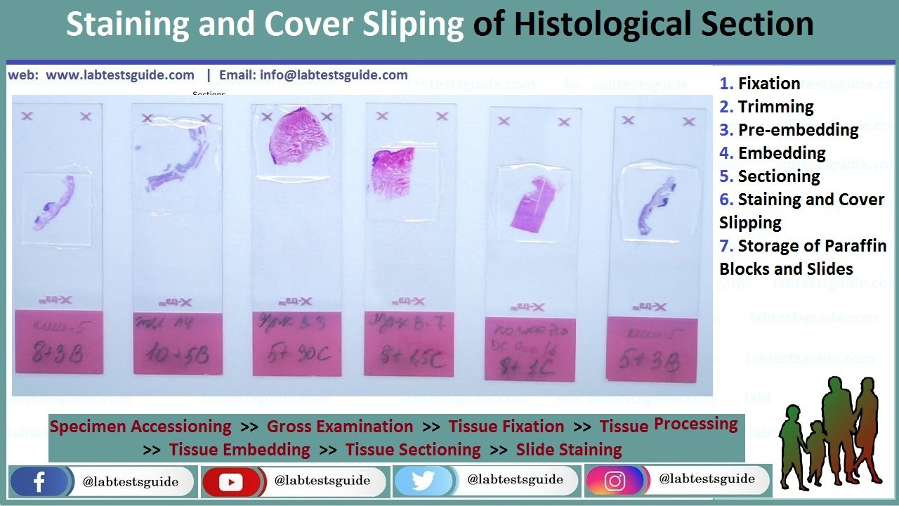
The Bitesize Guide to Special Stains for Histology Contents 2.

. Early histologists used the readily available chemicals to prepare tissues for microscopic studies. Fixation Fixation is the addition of special substances such as chemicals to tissues under investigation to preserve them by halting the progression of various biochemical processes that lead to degradation 1. Current used histological stains appear to be economical quick and reliable tools for interpreting archiving and delivering essential diagnoses that could not be achieved by any other means.
Carmine hematoxylin silver nitrate Giemsa trichome stain Gram stain and mauveine were among the first histological stains discovered in nature. Stains for carbohydrates 2. National Center for Biotechnology Information.
These laboratory chemicals were potassium dichromate alcohol and the mercuric chloride to harden cellular tissues. Salts - dissociate in aqueous solutions to form two ions -one ion is colored and can be either. - It is the preliminary or the first stain applied to the tissue sections - Gives diagnostic information in most cases.
A huge range of stains is used in histology from dyes and metals to labeled antibodies. A new deep-learning-based framework that generates virtually stained images using label-free tissue images in which different stains are merged following a micro-structure map defined by the user which could allow pathologists to get more relevant information from tissue and thus improve diagnoses. Guide for Histological Stains.
Histological staining is a series of technique processes undertaken in the preparation of sample tissues by staining using histological stains to aid in the microscope study Anderson 2011. Chemical reactions are also used to show up specific tissue components in special cases. These stain tissues in the same way they dye cloth.
For acidic dyes the dye in question can often in addition be selective for particular acidophilic components. Certain stains change the coloration of cells and tissues significantly different from the color of the original dye complex a phenomenon known as metachromasia. If used to visualize Australia Antigen HBsAg Hepatitis B Surface Antigen specific of hepatitis B virus the result must always be supported by immunohistochem- ical investigation.
Lacks Nissl bodies and does not stain with routine histological stains. Ganglion a collection of neuron cell. Histological staining is a vital step in diagnosing various diseases and has been used for more than a century to provide contrast in tissue sections rendering the tissue constituents visible for.
The process of histological staining involves five primary stages namely fixation processing embedding sectioning and staining. PDF UTILIZATION OF 1 OF METHYLENE BLUE IN STAINING HISTOPATHOLOGICAL PREPARATIONS AT ANATOMIC PATHOLOGY LABORATORY T. Histological and Histochemical Methods was first pub-lished in 1999 but it has been extensively updated for this edition by the addition of new procedures and the exten- sion of some of the chapters to include some older but useful techniques.
Gomoris Methenamine Silver GMS Stain for Microorganisms and Fungi 14. 269 What is pathology. The process of histological staining takes five key stages which involve.
Acid-Fast Stain for Microorganisms 11. Published September 6 2017 With the use of stains and dyes histology allows researchers to visualize particular tissue structures chemical elements within cells tissues and even microorganisms. A special stain is a staining technique to highlight various individual tissue component once we have preliminary information from the HE stain.
Toluidine Blue Stain for Mast Cells Cancer Screening and Forensics 16. Histological staining is a vital step in diagnosing various. Aside from their utility in.
Gram Staining for Bacteria 7. Axons are either myelinated surrounded by a fatty insulating sheath that speeds conduction of the electrical impulse or non-myelinated lacking a myelin sheath and thus conduct impulses slowly. Peter Michalka Lucia Donárová Ústav patologickej anatómie LFUK a UN pracovisko Staré mesto Sasinkova 4 Bratislava Prof.
HiStologiCal StaiNiNg Kit 12 13 ACID oRCEIN code 010251 Description The Kit is intended for use in histological visualization of elastic fibers with acid orcein. For basic dyes the reaction of the anionic groups of cells these include the phosphate groups of nucleic acids sulphate groups of glycosoaminoglycans and carboxyl groups of proteins depends on the pH at which they are used. Staining techniques used were carmine silver nitrate Giemsa Trichrome Stains Gram Stain and Hematoxylin among others.
The advent and evolution of histology follows that of microscopy as outlined in A very Short History of Histology. Processing biological material tissue samples from living or dead. SPECIAL STAINS IN HISTOLOGY STAINS FOR MICROORGANISM CONNECTIVE TISSUE STAINS STAINS FOR PIGMENTS AND MINERAL INTRODUCTION Most infectious agents are rendered harmless by direct exposure to formal saline fixative.
HISTOLOGICAL STAINS AND SOME HINTS FOR ANALYZING SLIDES 1 STAINS Many stains in histology were adapted from textile dyes. In general there is more discussion about the fixation processing and staining of microorgan-isms and plants. HE stains may stain many organisms.
Fixation processing embedding sectioning and staining Titford 2009. Histological staining acid basic histochemistry. Standard fixation process should be sufficient to kill microorganisms.
For staining paraffin sections of tissue are normally used. Warthin-Starry Stain for Microorganisms 9.
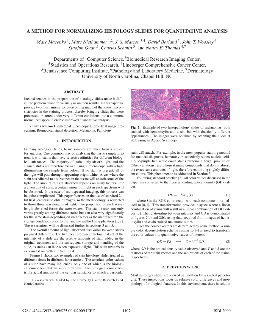
Pdf A Method For Normalizing Histology Slides For Quantitative Analysis

Pdf Computational Histological Staining And Destaining Of Prostate Core Biopsy Rgb Images With Generative Adversarial Neural Networks Semantic Scholar

Special Stains In Histopathological Techniques Pdf 1 Staining Of Carbohydrates Periodic Acid Schiff Pas Pas With Diastase Best Carmine Langhan S Course Hero

Pdf Introduction To Special Stains Semantic Scholar
.jpg)
Eosin Stains For Histology
Staining And Cover Slipping Lab Tests Guide

Pdf Introduction To Special Stains Semantic Scholar

Pdf Computational Histological Staining And Destaining Of Prostate Core Biopsy Rgb Images With Generative Adversarial Neural Networks Semantic Scholar
Staining Histology Cytology Histology Cytology Pathology Products
.jpg)
Hematoxylin Stains For Histology
Pdf Histological Stains A Literature Review And Case Study

Pdf Comparison Of Special Stains For Keratin With Routine Hematoxylin And Eosin Stain Semantic Scholar

Pas Stain Histology
Pdf Introduction To Special Stains Semantic Scholar
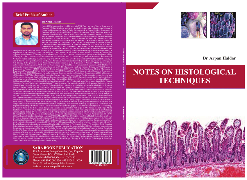
Pdf Notes On Histological Techniques
Pdf Dyes And Stains From Molecular Structure To Histological Application
Pdf Basic Histological Techniques For Planarians
Pdf Staining Efficacy Of Rose Extract In Comparison With Eosin Stain A Histological Study On Oral Tissues

Pdf A Study Of Xylene Free Hematoxylin And Eosin Staining Procedure