
One thousand forty-five OPGs were randomly chosen from patient population. Incisive canal cysts are treated with complete surgical removal by a palatal approach with the palatal flap.
Radiographic Appearance Of Cysts Part 3 And Scintigraphy Intelligent Dental
These cysts have no direct relationship to the teeth but in their growth may encroach upon the incisor apices.

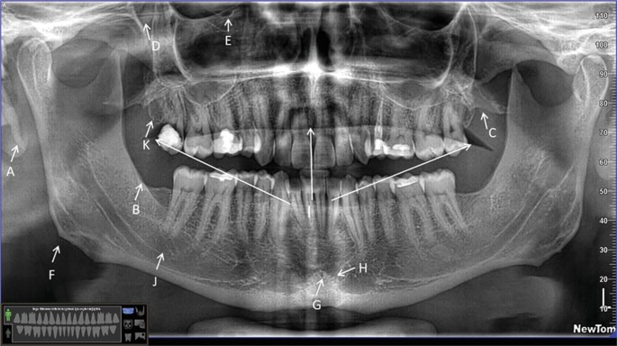
. Assessment of the mandibular incisive canal by panoramic radiograph and cone-beam computed tomography. The persistence of ductal epithelium leads to formation of cyst. Panoramic radiographs can be used for visualization of the mental foramen and a potential anterior looping but not for locating the mandibular incisive.
Our goal is to evaluate identification of MIC by both panoramic radiograph PAN and cone-beam computed tomography CBCT. According to Shear and Speight7 a radiographic shadow with antero-posterior dimensions of as much as 10 mm in the incisive fossa region may be. Incisive canal cysts also known as nasopalatine duct cysts NPDC are developmental non-neoplastic cysts arising from degeneration of nasopalatine ducts.
Cone-beam computerized tomography CBCT can show an uncommon mandibular incisive canal that cannot be detected by panoramic radiography which is used preoperatively to form the initial plan of the size and length of an implant fixture for surgical placement in the mandibular interforaminal area. The incisive foramen is the inferior opening of the nasopalatine canal incisive canal. The nasopalatine duct cyst occurs in the nasopalatine or incisive canal and it may be difficult to decide on a radiograph whether radiolucency in that area is a cyst or a large incisive foramen.
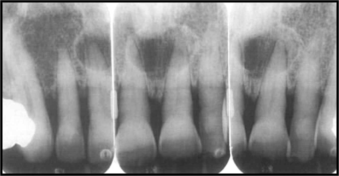
The incisive foramen also known as nasopalatine foramen or anterior palatine foramen is the oral opening of the nasopalatine canalIt is located in the maxilla in the incisive fossa midline in the palate posterior to the central incisors at the junction of the medial palatine and incisive sutures. NASOPALATINE duct cysts are cysts which form in the incisor canal region of the maxilla and originate in the nasopalatine duct or its remnants. The risk of damaging the mandibular incisive canal MIC during surgery in the anterior mandible should not be overlooked.
It can be single or multiple. The region between mental foramens is considered as a zone of choice for implants. Up to 10 cash back An incisive canal was identified in 15 of the images with good visibility in only 1.
The mandibular canal also known as the inferior alveolar canal IAC is located within the internal aspect of the mandible and contains the inferior alveolar nerve artery and vein. Determination of the position and anatomical. Panoramic radiographs can be used for visualization of the mental foramen and a potential anterior looping but not for locating the mandibular incisive.
An incisive canal was identified in 15 of the images with good visibility in only 1. Periapical radiograph panoramic and CBCT are needed to assess the lesion with better precision and limits radiation. Today I thought Id go with a little refresher of foramina visible on intraoral radiographs specifically periapical radiographs.
The incisive foramen generally appears in most panoramic radiographs though not with the clarity seen in periapical radiographs. It transmits the greater palatine artery and. However complications may arise due to an extension anterior to the mental foramen that forms the mandible incisive canal MIC.
Our goal is to evaluate identification of MIC by both panoramic radiograph PAN and cone-beam computed tomography CBCT. An incisive canal was identified in 15 of the images with good visibility in only 1. These ducts usually regress in fetal life.
The nasopalatine canal presents as a vertical radiolucent band between the roots of the maxillary central incisors superiorly to. There will be a total of 4 Im covering 2 each in the maxilla and mandible. Outline of the cyst.
After radiographic examination the buccal cortical plate of the mandible was remov. Hence preoperative radiographic assessment is essential to avoid complications. An anatomical variation to be considered is the anterior looping of the mental nerve in 11 of images.
An anatomical variation to be considered is the. It starts at the mandibular foramen on the lingual side of the ramus continues on the buccal surface of the mandibular body and ends at the mental foramen adjacent to the second. It is seen on both intraoral radiographs and extraoral radiographs.
The aim of this study was to investigate the presence of a MIC in panoramic radiographs OPGs. It is considered the most common non-odontogenic cyst and develops only in the. Although occasionally observed in radiographic examinations of the incisor area of the maxilla nasopalatine duct cysts were.
An incisive canal cyst is a developmental cyst non neoplastic cyst arising from degeneration of nasopalatine ducts. These ducts usually regress in fetal life. The study group comprised of 46 hemimandibles fixed in formalin.
However complications may arise due to an extension anterior to the mental foramen that forms the mandible incisive canal MIC. The pear-shaped radiolucency between the apices of the central incisors can be mistaken for periapical pathology or cyst formation. Only in a very few radiographs will the incisive canal or nasopalatine canal be.
The mandibular incisive canal MIC is the anterior extension of the mandibular canal and its presence is of interest in surgical procedures in the chin region. The differential diagnosis for incisive canal cyst includes medial enlarged nasopalatine duct central giant cell. Its time for the next canal.
This canal may also be referred to as the incisive canal. The region between mental foramens is considered as a zone of choice for implants. An anatomical variation to be considered is the anterior looping of the mental nerve in 11 of images.
So onto the maxilla first. The purpose of this article was to define the anatomic and radiographic courses of the incisive mandibular canal and discuss its clinical significance. They are seen as a solitary well-defined oval or round unilocular radiolucency between central incisors 6 mm in.

Intraoral Radiographs Identifying Normal Anatomy Today S Veterinary Practice

Panoramic Radiograph Image A Axial B And Oblique Sagittal C Ct Download Scientific Diagram
Panoramic Radiographic Anatomy Springerlink

Maxillary Anterior Landmarks Intraoral Radiographic Anatomy Continuing Education Course Dentalcare Com

Figure 2 Assessment Of The Mandibular Incisive Canal By Panoramic Radiograph And Cone Beam Computed Tomography

Anatomical Landmarks Of Panoramic Radiographs With Ppt Lecture Note For Download Lecture Notes In Dental Assistant Study Dental Hygiene School Dentistry

Opg Showing Incisive Foramen And Mental Foramen Download Scientific Diagram

Mouth Incisive Canal Cyst Professional Radiology Outcomes

Dentaltown Where The Dental Community Lives Denti Dentista Odontoiatria

Maxillary Anterior Landmarks Intraoral Radiographic Anatomy Continuing Education Course Dentalcare Com

Visibility Of Mandibular Anatomical Landmarks In Panoramic Radiography A Retrospective Study Semantic Scholar

Dentistry Lectures For Mfds Mjdf Nbde Ore Anatomical Landmarks Of Panoramic Radiographs With Ppt Lecture Note Panoramic Radiograph Dentistry Radiographer

The Radiographic Examination Intraoral Radiographs Of Maxillary Download Scientific Diagram

Periapical Radiograph 1 Year After Treatment Bone And Teeth Showing Download Scientific Diagram

Intraoral Radiographs Identifying Normal Anatomy Today S Veterinary Practice

Maxillary Anterior Landmarks Intraoral Radiographic Anatomy Continuing Education Course Dentalcare Com

Route Of The Incisive Canal Of The Mandible Mic Download Scientific Diagram

Maxillary Anterior Landmarks Intraoral Radiographic Anatomy Continuing Education Course Dentalcare Com

The Radiology Of Developmental Dental Defects Demystified An E Based Learning System Intechopen